Figure 1. Relative gain of measured parameters (age,
mass, size) between I and II exam as a tangent of an angle of different
fetal maturation rates (F - fast, A - average, S - slow)
M.F. Fathalla
On the initiative of the Polish Gynecologists Society, an international conference “The Childbirth in the Individual Term with the Use of the New Technologies” was organized in Cracow on 6th April 2001 in consultation with the FIGO President Shirish S. Sheth. Amongst the participants of the Conference were P.T. Professors and the heads of the University Ob/Gyn Departments from the Czech Republic (Z.Hájek from Prague, A. Roztočil, Z.Maly and V.Unzeitig from Brno), the Slovak Republic (J. Štencl from Bratislava, S.Lukačín from Košice, J.Dubčák from Dolny Kubin), Sweden (P. Fedor-Freybergh) and Poland (A.Basta, M.Klimek, R.Klimek, J. Krzysiek, R.Lauterbach, A.Reroń, A.Skręt and Z.Zdebski from Cracow, G.H.Bręborowicz, T.Pisarski and S.Sajdak from Poznań, B. Chazan, K. Czajkowski, R.Dębski and L.Marianowski from Warsaw, K.Kamiński from Katowice, A. Malarewicz from Kielce, W. Szymański from Bydgoszcz, J.Oleszczuk from Lublin and J. Wilczyński from Łódź).
The participants familiarized themselves in advance with the content of five papers posted in the Internet (http://www.ginekologia.info.pl): “Let a man be born at his own due time” by R.Klimek, “Prediction of the birth term and course of the labor” by M.Klimek, “Postnatal clinical assessment of fetal maturity in newborn infants” by R. Lauterbach, “Operative deliveries, professional and economic aspects” by Z.Hájek, L. Haaková and D. Kolarik and “The Contribution of Continual Fetal Oxygen Saturation (FspO2) by Means of Intrapartum Fetal Pulse Oximetry (IFPO) to the Diagnosis of Fetal Hypoxia” by A. Roztočil, M.Kučera, P.Kachlik, J.Miklica, P.Ventruba. Those papers were discussed in Cracow and served as the basis for formulation of the Declaration of the Childbirth in XXI Century [1] which is to be presented to the FIGO Committee of Ethical Aspects of Human Reproduction and Women’s Health for discussion and circulation according to the competence of this Committee. All Conference materials are available at the above mentioned Internet address and are to be published in Archives of Perinatal Medicine.
Our aim is to force an industry and editors of newspapers to understand properly the relativity of pregnancy duration and thus to reduce the number of instrumental deliveries and prematurity rate according to the following sentences published in the book "A Time to Be Born" [2].
Gynecologists have mustered technologies that can save the lives and improve the health of women and their children. We can cure diseases and relieve sufferings to an extent never known before. The impact on the health of women at large, however, still leaves much to be desired. (M.F. Fathalla)
Instrumental delivery has become a much too common form of birth in too many places, and pregnant women must suffer the consequences. It is time, therefore, to reevaluate current obstetrical practices and the attitudes towards these practices. (E.V. Cosmi, R. Klimek)
Every baby should be born a healthy baby. (J.J. Sciarra)
References:
Declaration of the Childbirth in XXI Century
R.Klimek (Ed.)*, A.Basta (Cracow), G.H.Bręborowicz (Poznań), B. Chazan (Warsaw), K. Czajkowski (Warsaw), R.Dębski (Warsaw), J.Dubčák (Dolny Kubin), P.Fedor-Freybergh (Stockholm), Z.Hájek (Prague), K.Kamiński (Katowice), M.Klimek (Cracow), J.Krzysiek (Cracow), R.Lauterbach (Cracow), S.Lukačín (Košice), A.Malarewicz (Kielce), Z.Maly (Brno), L.Marianowski (Warsaw), J.Oleszczuk (Lublin), T.Pisarski (Poznań), A.Reroń (Cracow), A.Roztočil (Brno), S.Sajdak (Poznań), A.Skręt (Cracow), J.Štencl (Bratislava), W.Szymański (Bydgoszcz), J.Wilczyński (Łódź), V.Unzeitig (Brno), Z.Zdebski (Cracow)
Introduction
The basis of each medical activity is the perennial rule Primum non nocere and only subsequently nursing, diagnosing, treatment and rehabilitation of the man who by himself alone is not able to secure sufficient help in health and disease due to lack of knowledge, skills and/or objective environmental conditions. In that light, gynecologists have a particular role to play because in helping women they accompany human beings from the conception. It has to be stressed that according to the modern knowledge the state of the mother’s health is of crucial importance as it is the natural environment in the fetal period of man’s life.
Pregnancy is the best example of the importance of environment in existence and development of every system. According to the quantum thermodynamics, they exchange matter and energy with each other through the system’s edges, which is accompanied by an increase of the general entropy. From the beginning of the pregnancy, the maturing fetus owing to its improving structure and operation quickly diminishes production of its own entropy and its exchange with the mother’s organism, while at the same time increasing the mutual material and energy exchanges. Owing to this, during the rapid change of its environment during the labor it is easier for the newborn baby to maintain independent exchange of entropy and energy with the external world. The introduction of the spectroscopy and NMR imaging in medicine enables obiectivization and measurement of the each event which occurs on the level of atoms and molecules, which always are the starting point of processes, which in the nearest time will take the form of chemical, biological and then medical phenomena.
Reaching full fetal maturity by the child coincides with the end of the biological existence of placenta and with the optimal internal state of the mother for the delivery. This modern interpretation of the cause of the initiation of labor is a subject of study for medical quantum thermodynamics, which is the basis of multifactoral physical, chemical, biological and psycho-emotional conditionings of the pregnancy. Therefore it is necessary to realize both this concurrence and diversity of changes which determine the individual birth so as not to rush into decisions about delivery without taking heed of individual fetal development and to contradict the words of the Polish poet Jan Twardowski: “To be born is a greater risk than to die” from his poem “Let us hurry to love people; they depart so quickly”.
Owing to the quantization of the fetal maturation process, one can use the evaluation of the increase in fetal spatial dimensions (by means of generally available ultrasound devices) to determine accurately the date of individual birth. Thus, a discussion about entropy or auxology is no longer utopian, and becomes a modern explanation of the perennial event. Directly after the delivery we can clinically verify the accuracy of the birth prognosis done with the help of modern ultrasound devices which were constructed on the basis of quantum thermodynamics. Therefore we recommend the introduction of ten rules of our declaration into clinical practice.
Declaration
1.The distribution of births in singleton human pregnancies is normal and consistent with the Gaussian curve, with an average duration of 281+/- 11 days (range 259-303 days), modal 283 and median 282 from the last menstrual period, which was proven on several millions of prospectively observed pregnancies. [1,5,11,12,16,24,29,30,33, 38,42,47,58,65,76] It follows that:
10 Approximately 60% of fetuses mature regularly until the 390/7 - 406/7 birth week, 20% mature fast and 20% mature slowly. Only 2.5% of deliveries occur before the 259th day and 2.5% after 303rd day.
20 Less than 4% of children are born on the middle day as defined by Neagele’s rule, while 3% are born in the extreme weeks of the 6-week norm.
30 97.5% of the children born before the 259th day (before 370/7 week) are immature but only 2.5% of premature deliveries occur in the range of the 390/7 – 432/7 weeks.
40 An average duration of the twin pregnancy which accounts for 1-2% of all deliveries is 259+/- 22 days which means that 50% of them are born in the first half of the period of norm for singleton pregnancies.
2.Labor occurs when the fetus reaches maturity to independent life which is the basic criterion of its proper starting point in physiological conditions. Mature fetuses can differ significantly in mass (mean 3 400+/- 400g, range 2 600-4 200g) and body length (54+/- 3cm, range 48-60cm) which show high positive correlation (r=0.75) with each other. The fetuses show positive correlation with the gestational age (r=0.32) on the same level, as with mass (r=0.3) and length (r=0.26) of the fetal body (p< 0.001).
3. Calendar time is not directly connected with either the process of fetal maturation or the fetal structure, and only registers sequences of events e.g. the date of the last menstrual period or successive ultrasonographic measurements. Therefore it cannot be the sole basis of clinical procedures as it does not concern the specific individual pregnancy but average statistical data. The claim that each pregnancy > 36 weeks is already mature is true only in 1-2% of cases because e.g. more than half of those babies are born after the Naegele’s term. And reversely, pregnancy must not be recognized as post-term just because the 41st week has passed on the calendar scale, when only from the statistical point of view several percent of fetuses still have to mature. Labor initiated at improper calendar time not only is usually prolonged and too often instrumentally terminated but also has negative consequences both medical and socio-economical. The threshold of 10% instrumental deliveries should be a significant dividing line of good perinatological care.
4. All ultrasound devices in the world, although built on the basis of the most modern knowledge, have biometric scales which use only calendar time. [10,12,27,34] Also their average values do not cover the fetal examined parameters >= 40 weeks or >= 41 weeks. Thus at least more than fifteen percent of normally developing fetuses remain out of ultrasound observation. In contemporary clinical practice even the most sophisticated ultrasonographic imaging equipment fails to provide in advanced pregnancy more accurate information relating to individual birth date than Naegele's Rule (95% confidence interval ± 3 weeks) using only the knowledge of the LMP date. In 1985 S.Campbell and co-workers [2] verified Naegele's Rule on 4 000 BPD measurements though their ultrasonographic interpretation contained a cardinal error committed, unfortunately, as recently as 1994 [9]. The error lies not in the technologically superior equipment but rather in the application of its programs. Their authors so far have failed to react to critical publications that appeared to date [11,36,37,39,40,43,44,58]. For example, the 400/7-6/7 week on the calendar scale may represent for all observed babies only one of three different possibilities: 1) the 40th week for the babies in which delivery is to occur (i.e. birth only for 30% cases), but 2) some babies to be delivered during the 37th week are actually three weeks overdue, while 3) those to be delivered during the 42nd week still have two more weeks prior to birth. Therefore pregnancy dating scale which is incorporated in USG programs [7,8,14,27,64,70,71] can only provide a statistically probable range of delivery since calendar time (called horizontal time due to its linear appearance) is not directly connected to the fetuses' rate of maturity [19,21,23,25,27,44,58].
The object of this declaration is to draw attention to the responsibility of industry for programming their products, if their use could entail medically harmful effects. Instrument, which is advertised as the one that enables to asses fetal age, must not obtain data differing by 1 or 2 weeks in the average values of pregnancy duration, depending on which scale was chosen by the manufacturers (Tables 1-4). And reversibly, a physician who observes closely the development of pregnancy from the last menstrual period, will not acquire data for e.g. 420/7 week, because some scales end at week 40 or 41.
Table 1. Mean values of BPD (mm) at the same gestational
age (weeks) according to Asia (I, II, III) and Europe (IV, V, VI) scales
|
|
|
|||||
|
|
|
|
|
|
|
|
|
|
|
|
|
|
|
|
|
|
|
|
|
|
|
|
|
|
|
|
|
|
|
|
Table 2. Mean values of FL (mm) at the same gestational
age (weeks) according to Asia (I, II, III) and Europe (IV, V, VI) scales
|
|
|
||||
|
|
|
|
|
|
|
|
|
|
|
|
|
|
|
|
|
|
|
|
|
|
|
|
|
|
|
|
Table 3. Mean gestational age indicated by Asia (I, II,
III) and Europe (IV, V, VI) BPD scales
|
|
|
|||||
|
|
|
|
|
|
|
|
|
|
|
|
|
|
|
|
|
|
|
|
|
|
|
|
|
|
|
|
|
|
|
|
Table 4. Mean gestational age indicated by Asia (I, II,
III) and Europe (IV, V, VI) FL scales
|
|
|
||||
|
|
|
|
|
|
|
|
|
|
|
|
|
|
|
|
|
|
|
|
|
|
|
|
|
|
|
|
Birth is both the most important, though also one of the most dangerous moments because of its potential consequences in every person's life. Improper scales found in ultrasonographic equipment obstruct the use of this wonderful apparatus in birth prognosis and first of all lead to unnecessary blood loss by thousands of women undergoing instrumental deliveries in the most industrialized countries of the world. If such births cause but an average of more 100ml blood loss during each delivery then their sum total of useless loss must call to mind the tragedy of I.F.Semmelweis and G. Michaelis and the conservatism of their opponents. Therein lies the essence of personal or industrial responsibility in various existing birth scales. However, it is most important, to entrust this task to the editors of textbooks and obstetrical journals whose authors still publish data mistaking the notion of pregnancy weeks and birth week as well as attaching greater importance to the beginning of pregnancy than to its more important goal which is, of course, its successful conclusion.
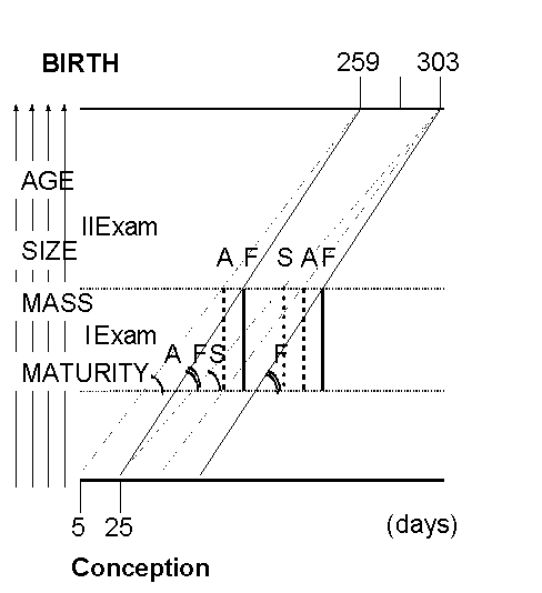
5. Fetal maturation is a timespatial process which means that from the increase of spatial parameters one can calculate the individual time of maturation expressed in the number of technical quanta. [20,21,23] The rate of their appearing determines the birth term independently of the last menstrual period or in vitro fertilization! Maturity quanta are measured on the same axis as mass, length or individual values of biometric ultrasonographic measurements. Making two measurements in the advanced pregnancy (>= 28 weeks) at the interval of >= 2 weeks, one can not only determine the current condition and fetal age but also birth term and predicted maturity, mass and length of newborn-to-be. [13,22,25,42,48,56]
6. Biometric scales of few fetal parameters used in ultrasound devices are not correlated with one another therefore the result that assessment of fetal age can differ even in average values by 2 weeks depending on the examined parameter e.g. femur length, head or cranium circumference. It is caused by the fact that it is not the absolute value of the examined parameters and the postmenstrual pregnancy duration which determine the birth term and the condition of the newborns but the constant growth rate of all spatial fetal parameters. This is completely ignored by the manufacturers of ultrasound devices in spite of repeated protests of the FIGO Study Group for Assessment of New Technology [12,34] and addresses at World Congresses of FIGO, Ultrasonography and Perinatology [4,26,27,35,39,41, 43,45,46,56,57,62,68].
7. The quantum thermodynamics makes possible prediction of the birth term with the accuracy of +/- 3-4 days. Owing to this we consider premature each end of pregnancy which occurs >= 1 week before, and post-term if the labor does not occur >= 1 week after the individual term. In each of three components of pregnancy (fetus, mother and placenta with the fetal membranes) several days before the birth term there occur clinically noticeable changes which enable verification of the clinical prognoses of the date and course of labor. Diagnostics at the last week of pregnancy is particularly important, especially that of uterus reactivity before the pharmacological induction of labor even when treated as a functional test. [21,47,51]
8. Adjustment of the existing ultrasound devices to the requirements of the modern medical thermodynamics was patented first in Poland and later in Europe (European Patent No 0 557 831) which not only proves the originality of the method of ultrasonographic fetal biometry and birth prognosis with the accuracy of several days but should primarily force the manufacturers and designers of the faulty scales to change forthwith their way of doing things due to the growing hazard for human health.
Neonates that grow fast or slowly in pregnancy have at birth the same
number of quanta of maturity although the frequency of appearance of their
smallest portions of maturity (i.e. quantum) is different. So, by performing
two ultrasonographic measurements one can in addition to measuring growth
itself, simultaneously evaluate its rate. The same absolute increase of
measured ultrasonic parameters between two serial measurements do not have
a distinctive quality, but decisive is the angle at which growth line will
take place over a known period of time [19,21-23]. Figure 1 shows how to
designate the already mentioned angle to growth. Under physiological conditions
all tested parameters have the same rate of maturity.
Figure 1. Relative gain of measured parameters (age,
mass, size) between I and II exam as a tangent of an angle of different
fetal maturation rates (F - fast, A - average, S - slow)
Technological advances do not always go hand in hand with adequate improvement of therapeutic results, and at times even lead to early and delayed iatrogenic effects, as in the case of dating pregnancy duration with the help of ultrasonographic devices. In the area of obstetrical ultrasonography, the physicians' attempts to solve this problem have failed as yet due to the manufacturers' reluctance or inability (for technical and economic reason) to change the inaccurate and clinically harmful software, which is responsible for the unethical increase in the number of instrumental deliveries.
9. Individual evaluation of each labor ought to be performed directly after the delivery by routine assessment of the adaptation of the newborn baby in Apgar scale and of its maturation according to Klimek index. [36,50,59] On the basis of examination of 3 662 newborns the maturity index K was calculated as 9+/- 1.5 points. There was found a high correlation (r=0.69, p<0.01) between the new index and the Ballard scale. The new index K is simple and produces comparable resultes encompassing a full maturiy in the range of only 6-12 points It is particularly important in the case of instrumental deliveries. The condition of the newborn depends mainly on its gestational environment and thus testifies to its mother’s health. This is what differentiates modern medical thermodynamics from the labor mechanics of the bygone era of the absolute time and space. Medicine which is focused only on pathology does not fulfill the requirements of the modern perinatology. which points to the necessity of using the entire human knowledge and does not allow to change physiological events into pathological ones. Both the prophylaxis of premature labors (which belongs to pathology) and non-interference with natural gestational processes (physiology) through unnecessary labor induction or cesarean section at a time improper for individual pregnancy, are more important than treatment of premature infants with maturity index K<6 points.
Table 5. Scoring system for simplified clinical assessment of Klimek’s maturation index in newborn infants
|
|
|
|
|
|
|
 |
 |
 |
|
To arm |

100o-180o |

90o-100o |

<90o |
|
|
 |
 |
|
|
|
|
|
|
|
|
|
|
|
|
|
bud<3 mm |
bud 3-4 mm |
Bud > 4mm |
10. Irrespective of the need to modernize ultrasonographic scales also enzymatic monitoring of pregnancy has to be revitalized. As far back as 1930 it has been demonstrated by K. Fekete that the serum of pregnant women was capable of abolishing the uterus-stimulating activity of posterior pituitary extracts. In late forties this feature was linked with enzymes activity called oxytocinase (E. W. Page, K. Semm, E. Werle, G. Effkeman, A. Hevelke, K. Buthmann), but the obtained results have not been adapted in clinical practice. In 1957 H. Tuppy and H. Nesvadba described the principles of chemical assay of the enzyme. They used as substrate L-cystine-di-betha-naphtylamide indicating erroneously a 1% concentration of solution of sodium nitrate instead of 0,1% what precluded the determination of oxytocinase. In 1961 R. Klimek and M. Pietrzycka revised the methodical error and made the first attempt at establishing an international unit of enzyme activity: umol/l/min. [61]
The following years brought an increasing interest in assessment of aminopeptidase activity in obstetrics (B. Berde, J. Berhard, P. Fylling, G. Graff, T. Hashimoto, H. Kleiner, R. Klimek, S. E. H. Melander. Z. B. Miller, W. Muller-Hartburg, A. P. M. van Oudeusden, A. M. Riad, A.M. Rutenburg, G. Ryden, S. Sakamoto, I. Sjoholm, E.E. Smith, F. Sorm, M. A. Titus, P. Wilken et al.). The European especially Scandinavian authors were leading ones in this field. Unfortunately in 1966 C. Babuna and E. Yenen published a wrong study on the modification of the original chemical assessment of oxytocinase [2,3], which in fact hindered investigations of enzyme monitoring in pregnancy. Three years later R.Klimek delivered a paper in which these laboratory mistakes were corrected. [60] Due to refusing its publication in American Journal of Obstetrics and Gynecology ( although this journal published his other clinical studies [6,53,54]) it was published in a biochemical journal which was readily available for the clinicians. Finally, the introduction of new methods of pregnancy monitoring, e. g. human placental lactogen, sonography as well as divergences of opinion on oxytocinase application resulted in groundless neglect of the study on cystine-aminopeptidase activity of pregnant women serum. The continuation of our investigation on the use of various substrates and buffers resulted in the clinical verification of chemical methods that could be applied in the monitoring of pregnancy for the prediction of delivery.
Oxytocinase and its isoenzymes reflect the present state of the mother, the child and the placenta – the three being inseparable until the labor components of pregnancy. Maternal blood levels of cystine-aminopeptidases (CAP1 and CAP2) show high correlation with the fetal and placental mass as well as the fetal maturity level.[6,28-32,49,63]
Maternal blood levels of cystine-aminopeptidases (CAP1 and CAP2) in the 4 consecutive weeks of the sixth gestational month were correlated with the fetal maturation rate. Then the constant increase of oxytocinase and isooxytocinase in maternal blood up to the time of delivery in 81% of pregnancies and potential stabilization of its level during the last two weeks of pregnancy in further 12% of cases are the most sensitive indicators of the proper development of fetuses. Enzymes levels and the rate of their increases enable to predict the probable birth term with all the more accuracy, the more similar the subsequent values of the enzymatic determinations. Their low level during pregnancy proves the hypothalamo-hypophyseal hypofunction and is a pathognomonic symptom in the habitual abortions caused by such insufficiency. Decrease in enzyme levels instead of the normal constant increase precedes by several weeks intrauterine death of the fetus which reaches 70% when untreated in such neuroendocrine gestoses. The substitutive ACTH-depot therapy makes possible to end every pregnancy successfully, but with statistically lower (p<0.01) values of CAP1 and CAP2 by 0.2 umol/l/min till the 23rd gestational week and later two times higher differences in CAP2 level and 5 times higher for CAP1 level.
The object of this summary is to highlight the importance of enzymatic monitoring which serves as the basis of the biochemical diagnostics in all fields of medicine except for obstetrics. This is particularly odd, as the first enzyme used in medicine was the placenta-produced “defensive pregnancy enzyme” discovered by E.Abderhalden.
Conclusions
Let a man be born at his own due time which has to be assessed with an accuracy of +/- 3-4 days according to the individual developmental time, not to the calendar scale which enables only the determination of its six-weeks range. According to the laws of statistics and auxology, new scales have to be introduced in ultrasound devices in order to bring the rate of instrumental deliveries (mainly cesarean sections) below the level 10% of all deliveries. Like in many other fields of medicine one has to restore enzymatic monitoring of pregnancy by the estimation of amnopeptidases (oxytocinase, cystaminopeptidase) in the mothers’ blood, which increase testifies to proper fetal development or if it is not the case anticipates fetal life hazard several weeks before the appearance of clinical symptoms. Fulfillment of those two postulates leads to a significant reduction not only of perinatal morbidity and mortality but also the costs of obstetrical care. The point is that only a part of the expenditure spared in this way suffices to introduce the postulated changes in ultrasound and enzymatic diagnostics (See: “Operative deliveries, professional and economic aspects” by Z.Hájek, L. Haaková i D. Kolarik and “The Contribution of Continual Fetal Oxygen Saturation (FspO2) by Means of Intrapartum Fetal Pulse Oximetry (IFPO) to the Diagnosis of Fetal Hypoxia” by A. Roztočil, M.Kučera, P.Kachlik, J.Miklica, P.Ventruba).
At the end of the seventies and beginning of the eighties, in countries with the most modern perinatal care, we have seen rapid growth in the indications for operative delivery with the aim of lowering perinatal mortality [67,73,74]. The international study of the WHO, ”Having a Baby in Europe,” [15] found increasing differences in the frequency of both caesarian sections and vaginal operative deliveries (forceps, vacuum extraction). This has been one of the most significant differences in obstetric care between the individual European countries [17,18]. However, with the increase in operative deliveries, there has not been a proportional decrease in perinatal mortality. The increase in expenses (longer hospital stay, maternal and neonatal complications) has also not been proportional to the benefits (decreased perinatal mortality and morbidity). [69,72,75]
Since 1995 in I Ob./Gyn Department of Charles University, Prague, there has been a rapid increase of caesarian sections. There has been almost an 8-fold increase in the number of caesarian sections, and the perinatal mortality has decreased 5.5 times. The number of vaginal obstetric operations (forceps deliveries) has remained constant between 2-3%. The greatest increase in the frequency of caesarian sections was noted in the last 5-6 years. The benefit of the increase in caesarian sections grows until reaching 10%, while further increase has no influence on lowering the perinatal mortality [67,74]. The increase in low- and high-risk states in pregnancy (vaginal delivery after a prior caesarian section, breech presentation delivery) plays a role in the gradual increase in caesarian sections [17,66]. Another factor influencing the frequency of caesarian sections is the obstetrician’s anxiety of a lawsuit in the case of neonatal death or disorders in the child. In the complicated cases, the constantly rising medical expenses negatively influence the hospital’s budget. In the Czech Republic, the frequency of complications after caesarian section is between 15-20% [73,74]. In 1998, the number of complications after caesarian section was 16.8%. The expenses for basic medical care have reached 713,000 CZK, when including intensive care in the cases with severe complications (coagulopathy, hysterectomy) the expenses are in the hundreds of thousands CZK. On a national level, the maternal mortality is alarming, although it is low at 0.11 per 1000, but in 61% of the cases is a association to cesarean section and in 29% is a direct relation to this operation.
One has to stress that the sole understanding of the rule of relativity of pregnancy duration and even realization of the naturalness of the fast, regular and slow fetal growth leads to the improvement of all indices of perinatal care. Nevertheless the proper attitude of the pregnant and potentially pregnant women is called for (See: “Let a man be born at his own due time” by R.Klimek, “Prediction of the birth term and course of the labor” by M.Klimek, “Postnatal clinical assessment of fetal maturity in newborn infants” by R. Lauterbach).
References: