“Let a man be born at his own
due time”
Ob/Gyn Departments Jagiellonian University
Kopernika 23, 31-501Cracow, Poland
Phone number: 0048 12 421 36 66
Quantum mechanics and theory of relativity are the most important scientific achievements of the twentieth century. Unfortunately, in spite of the great technological progress in the production of medical instruments and means of communication, they have not been used to the full in gynecological procedures. 100 years ago the notion of quantum and later timespace were introduced but obstetrician still adhere to out-of-date ideas of absolute time and space. As I. Newton in the seventeenth century they tend to put their trust in deterministic causality rather than their stochastic conditioning. To make matters worse, half of our colleagues - even if they know modern definitions of preterm and at term labor [23,24,29-31,40,41] – continue to assess fetal maturity according to fetal weight or calendar duration of pregnancy and on that premise they act improperly.
Pregnancy ends with reaching full fetal maturity, which can and has to be quantized like e.g. radiation and thermal or electric conduction. Likewise, mass is technically quantized in grams or kilograms, while gestational age is expressed in days, weeks or months. Maturity is neither mass nor time and cannot be assessed in grams or units of time. Therefore modern neonatologists and obstetricians successfully worked out maturity quantizing in points without taking into account mass, length or gestational age of fetuses and newborns [2,3,8,33-35,39]!
Science, Technology and Obstetrics
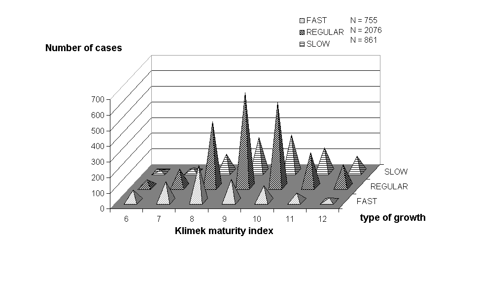
The fact that technologists install incorrect scales in ultrasonographic devices designed on the basis of the newest achievements of the twentieth century is reprehensible [5,7,25,27,28,30,36-38]. Everyone should be aware that on both sides of the mean value of the normal human pregnancy duration (281st-283rd day) there are two populations of newborns very much alike in their characteristics. Only 2.5% of these newborns have values of mass, length and maturity higher and lower than the range of the norm of the entire population, which pregnancies end within 6 weeks period. It was proven on hundreds of thousands and even millions (!) of observations of pregnancy duration in the whole world [1,4,10,42,44]! In the meantime, the scales of modern ultrasonographic devices do not cover the whole range of the 95% birth occurrence from 370/7 to 432/7 weeks after the last menstrual period. To make things worse, as the pregnancy progresses in weeks, the mean values of observed ultrasonographic measurements are falsely extrapolated or concealed by providing the wide range of standard duration +/- 3 weeks. These obvious arithmetic errors lead to iatrogenic and mediagenic diseases and deaths. Technicians not only use the falsely interpreted calendar scale of pregnancy durat ion playing up the role of the beginning of pregnancy at the expense of its much more important end. They entirely overlook the auxological laws, which are so well known from a later human developmental period i.e. sexual maturation. No one predicts the date of menarche on the basis of the absolute body mass or height but rather of the rate of their increase. Similarly, fetuses maturate slowly, regularly or fast, except that according to the auxological laws peers by the date conception but maturating faster have perinatal and target features smaller than the other children. Only newborns in 39th week have the distribution of their features closest to the normal. In fetuses which are born normally but earlier, the prevailing values are lower than the average, while in the most slowly maturating fetuses the situation is reverse.
One of the most frequent causes of instrumental deliveries, as well as the continuing 7-8% prematurity rate, is obstetricians ignorance of the achievements of auxology. Much better off are pediatricians, who observe auxological standards from the rate of birth of every human being. Every child up until the pubertal spurt generally develops in its own canal (range), whose change must be explained in every case. It is worthy of note that puberty is the next developmental stage following fetal maturation. Children which mature sooner have higher growth parameters than their peers, but the duration of their pubertal spurt is shorter and ultimately they grow to lower height. The same situation occurs before fetus reaches full maturity. The average values which determine weight and length of infants whose maturation is the fastest, i.e. those born in 37th week (37 0/7 - 37 6/7), are lower than in children born in 38th week, while it is only from 39th week that the characteristics of infants born in the following three labor weeks are stabilized. Only newborns in 39th week have the distribution of their features closest to the normal. In fetuses which are born normally but earlier, the prevailing values are lower than the average, while the most slowly maturing fetuses the situation is reverse (Figure 1).
Figure 1. Distribution of the neonatal features according to rate of fetal maturation
Thus, the auxological rules must not be disregarded. After all it suffices that in the proper period of pregnancy the obstetrician examines twice the development rate of fetal maturity. The data obtained from these examinations allow him to predict the birth-term with the accuracy of several days instead of weeks.
Real and statistical birth term
Everybody knows that he was born on a given day, while every obstetrician is able to prove that the commencing delivery and its prodromes occur during just two or three days of profound transformation from pregnancy to labor. Therefore the delivery date has to be estimated with accuracy of +/- 3 days. Unfortunately, to many obstetricians single out two days in the calendar pregnancy scale: the 259th day, starting from every fetus is magically considered “mature”, and then – somewhat contradictorily – the next day in question is 287th to 294th as the last one to determine maturity.
Extensive statistics show that from 287th day of the calendar scale to its 303rd day, there mature over ten percent of slowly developing fetuses. If the physician terminate these pregnancies by inducing labor, in at least several percent of cases he will deliver premature infants. If we consider 100 healthy women (100%) with the same beginning and normal course of pregnancy, 3 of them according to the Gauss curve will deliver in the first and 3 in the last week of the six-week range of the occurrence of births within 360/7 - 432/7 weeks after the last menstrual period, and 30 in each of the 2 middle weeks (390/7 - 406/7 ), while only 15 in 380/7 –386/7 and 410/7 –416/7 week. (Fig.2)
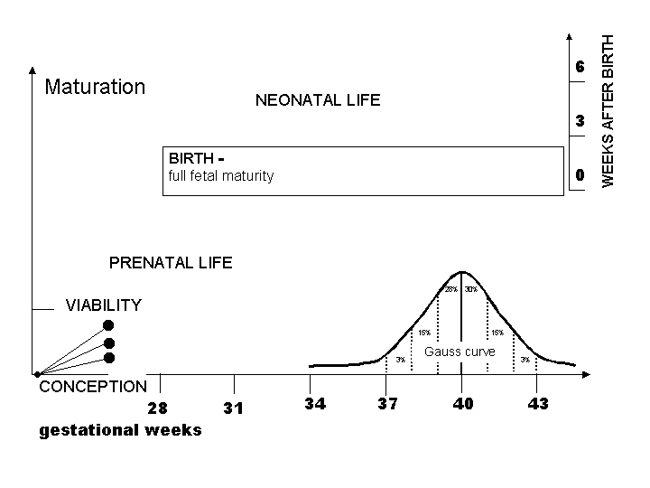
Figure 2. Normal occurence of human birth according to Gauss curve.
Pregnancy - just as every natural phenomenon (structure or process) - is an individual time spatial event whose most important element is fetal maturation of a human being, first to the level of viability, and then to full maturity to self-dependent life (Fig.3).

Figure 3 Fetal maturation to the level of viability and self-dependent life.
Out of the any group of 100 pregnant women with calendar age 370/7 - 376/7 weeks, only in 3 the process of fetal maturation is completed. Out of the remaining 97, only 15 will have it completed in 38th week and about twice as many in 39th week (Fig 4).
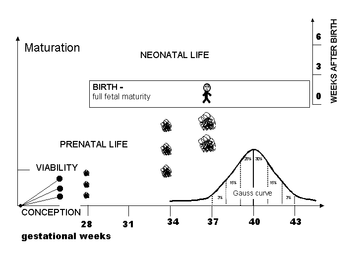
Figure 4 Full fetal maturity among 100 pregnant women at 370/7- 6/7 gestational week
When examine 50 pregnant women in 40th week, we have to realize that only approximately 30 out of their fetuses are mature for labor and other ones will deliver - unless pregnancy is iatrogenically terminated - after 287th day of physiologic pregnancy 410/7 week (Fig. 5).
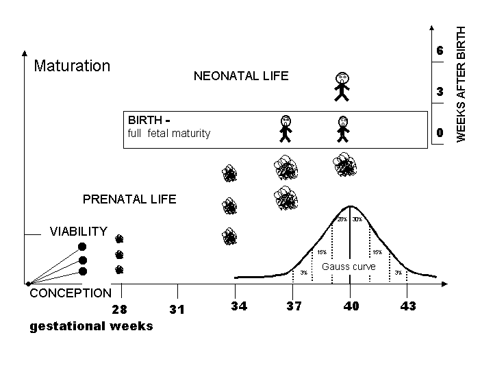
Figure 5 Full fetal maturity among 51 pregnant women in 400/7 - 6/7 gestational week
After the 42nd week still 3% of children can be physiologically delivered. (Fig.6)
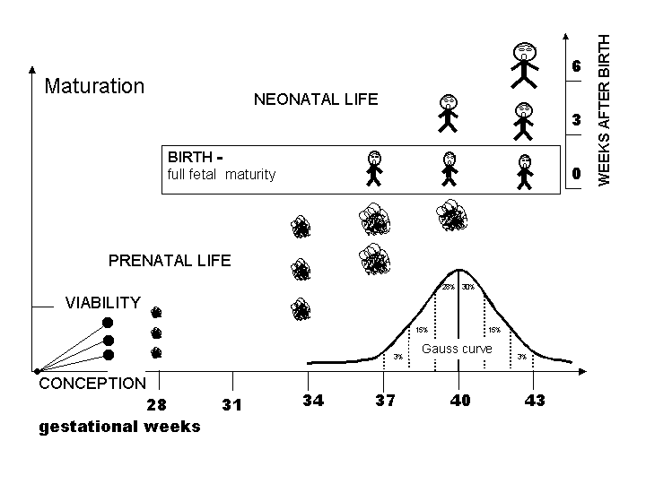
Figure 6 Comparison of pre-and postnatal ages of babies at 37th, 40th and 43rd weeks after LMP.
Conclusions
Modern medical means as ultrasonographic devices, cardiotocographs or neonatological incubators from the technical point of view stems from the greatest advances of quantum mechanics and theory of relativity. Unfortunately, their use in obstetrics paradoxically leads to iatrogenic morbidity and mortality due to lack of understanding of time-spatial fetal maturation and relativity of calendar pregnancy duration [6,11,14,22,26,30,32].
Labor occurred at an improper time is a common obstetrical error, which finds confirmation among others in higher perinatal mortality indexes both at the beginning (weeks 37th/38th) and the end (weeks 41st/42nd) of birth occurrence range in humans. The former is characterized by neglect of assistance in actual preterm labor one week before true individual term, and the latter by preterm labor induction or – even worse – attempts to bring belated assistance in postmature pregnancies, whose birth time has passed in the former weeks of the calendar scale of pregnancy duration.
By means of the presently used ultrasonographic devices but taking into account quantum mechanics and relativity of pregnancy duration one can - on the basis of two measurements made within 2 weeks - not only assess the current maturity, mass, length and gestational age of the unborn child but also predict those values in the perinatal period [13-22]. It brings measurable medical, social and financial profits and - most importantly – discards the ethics of reticence on the dangerous dominance of technology over general knowledge. It also reminds the doctors that their first obligation remains the tenet “primum non nocere”.
There was a time when improper use of cardiotocographs resulted in too much irreversible medical and social damage. The time has come to point one’s finger at manufacturers and users who bear the responsibility for similar effects in obstetrical ultrasonography. This is the best way to bring the percentage of prematurity down to the natural limit of 2.5% of all deliveries. Currently, 10-18% of labors are induced prematurely only because the calendar time of pregnancy duration has exceeded 287 or 294 days from the date of the last menstrual period, which additionally is given by the mothers accurate to several days, anyway.
The reduction of perinatal mortality – sometimes wrongly ascribed mainly to obstetricians – is primarily an effect of the amazing progress in neonatology. Low birth weight, perinatal mortality and prematurity rate have been even adopted as general social and economic indices of development of entire countries or at least selected territories. Therefore, to bring out the role of obstetricians there in, one should permanently introduce two other clinical criteria: distribution of birth in the range of six-week norm of occurrence in humans and ratio of premature infants to the mature ones at the gestational age <37 weeks counting from the last menstrual period.
References