The Contribution of Continual Fetal
Oxygen Saturation (FSpO2) by Means of Intrapartum Fetal Pulse
Oximetry (IFPO) to The Diagnosis of Acute Fetal Hypoxia
Fetal oxygen saturation role in diagnosis of acute hypoxia
1A. Roztočil, 1M.
Kučera, 2P. Kachlík, J. 1Miklica, 1P.
Ventruba
11st Deparetment of Gynecology and
Obstetrics, Medical Faculty, Masaryk University, Brno, Czech Republic
2 Deparetment of Special Education, Faculty
of Education, Masaryk University,
Brno, Czech Republic
Design: Open prospective study.
Setting: 1st Department of Gynecology and Obstetrics, Medical Faculty, Masaryk University, Brno, Czech Republic
Material and Methods: From January 1, 1999 to December 31, 2001 135 patients were enroled in the study. For the application of the IFPO sensor the patient had to meet the following criterias: patient´s informed consensus, pregnancy > or = 36 weeks, regular uterine contractions, rupture of membranes, cervical dilatation > or = 2 cm, singleton pregnancy, cephalic occiput presentation, no signs of vaginal infection and signs of acute fetal hypoxia on CTG tracing. We have used the fetal pulse oxymeter Nellcor Puritan Bennett N-400 and a Nellor FS 14 sensor. The sensor was aplied preferably on the posterior cheek of the fetus to all patients who fulfiled the above mentioned criteria in the 1st and 2nd stage of labor. The value of FSpO2 was continuously monitored up to complete dilatation. The treshold of intrapartum fetal hypoxia (FSpO2) was considered < 30 % for more than 10 min. In case of normal FSpO2 value the delivery was conducted vaginaly even if the CTG tracing continued to signalise intrauterine hypoxia. In case of patohologic FSpO2 value Caesarean Section was performed.
Results: IFPO in an easy feasible method. The FSpO2 value was obtained in all cases. There are no serious side efects neither on the mother nor on the fetus. No Caesarean section was performed in all patients with suspicious CTG tracing (35) after the verification of the FSpO2 value by means of IFPO. There were 100 patients with pathologic CTG tracing indicating the performance of Caesarean section. The Caesarean section was performed only in 27 patients after the verification of the FSpO2 value by means of IFPO. The remaining patients delivered vaginaly. There were statistical diference in the the FSpO2 value and in the postpartum umbilical cord pH between these two groups.
Conclusion: These results indicate that taking in an account fetal FSpO2 value evaluated by IFPO in the 1st stage of labor in case of pathologic CTG tracing indicating Caesarean Section lower the rate of Caearean Sections with identic perinatal outcome (Apgar Score, umbilical cord pH).
Key words: fetal oxygen saturation (FSpO2),
intrapartum fetal pulse oxymetry (IFPO), acute intrapartum fetal hypoxia,
Caesarean Section
CTG tracing is routine for monitoring fetal wellbeing antenatally. It is easy to carry out and being noninvasive has no serious side effects. To establish a diagnosis of acute intrapartum fetal hypoxia CTG tracing is provided whit high sensitivity but low specifity. This means that while all suspicious traces are noticed, not all of them are connected with acute intrapartum fetal hypoxia. According to literature sources the rate of the false positive traces is 40 - 70 %. Due to the fact that acute intrapartum fetal hypoxia is an indication for prompt delivery, due to over diagnosis from CTG tracing 40 - 70 % of labors end in emergency obstetric operations (Caesarean Section, forceps) eventhough acute intrapartum fetal hypoxia is not present.
Significantly, the frequency of Caesarean Sections is gradually increasing nowadays. The diagnosis of acute intrapartum fetal hypoxia is approximately 1/3 of cases of the indication for it. Worldwide, the trend is to decrease the number of Ceasarean Sections due to the notably higher rate of maternal mortality associated with it. To establish the diagnosis of acute intrapartum fetal hypoxia more precisely is one of the means by which Ceasarean Sections may be avoided.
There are two more methods of establishing the diagnosis of acute intrapartum fetal hypoxia more accurately.
1) Fetal blood sampling
2) Intrapartum fetal pulse oximetry (IFPO)
Fetal blood sampling to obtain a scalp blood pH value is an invasive method introduced into practise by Saling in the year 1967. It is often hard to carry out and its outcome may be influenced by hemodynamic changes in the scalp during the labor (caput succedaneum). Fetal blood sampling provides information about fetal wellbeing only at the time of bloodtaking. The main limitation of this type of examination is the necessity of taking further samples to update its results. Fetal blood sampling is usually used as an additional examination.
IFPO is a new method of direct and continual fetal oxygen saturation monitoring. It is a non invasive examination (apart from its involving the rupture of membranes and the intrauterine placing of the sensor) and does not have serious side effects. According to the literature IFPO is a complementary examination to CTG tracing used when it indicates the signs of suspicious acute intrapartum fetal hypoxia. Use of both CTG tracing and IFPO makes the diagnosis of acute intrapatrum fetal hypoxia more conclusive when only CTG tracing is used.
The aim of this study was to evalute the possibility of lowering the Caesarean Section rate in patients demonstrating the signs of acute intrapartum fetal hypoxia on CTG tracing.
Materials and methods
The study was carried out at the 1st Department of Gynecology and Obstetrics, Medical faculty, Masaryk University, Brno, Czech Republic from January 1, 1999 to December 31, 2000. 135 patients were enrolled in the open prospective study.
Criteria for enrolment:
- patient´s informed consensus
- pregnancy > or = 36 weeks
- regular uterine contractions
- rupture of membranes
- cervical dilatation of > or = 2 cm
- singleton pregnancy
- cephalic occiput presentation
- no signs of vaginal infection
- suspicious CTG tracing
- pathologic CTG tracing
Criteria for a suspicious CTG tracing:
- without accelerations in heart rate for more then 40 min.
- baseline heart rate between 150 to 170 beats/min and between 100 to
110 beats/min
with normal variability and without decelerations
- silent CTG tracing for longer than 40 minutes, normal base heart rate
without
decelerations
- variability > 25 beats/min. without accelerations in heart rate
- variable decelerations ( DIP 0 < 60 beats/min), > 60 sec.
- bradycardia < 80 beats/min. for > 2 min.
<100 beats/min for > 3 min.
Criteria for pathologic CTG tracing
- baseline heart rate < 100 beats/min.
- progressive bradycardia: baseline heart rate gradually decreases between
contraction
( DIP II, DIP 0)
- persisting bradycardia, baseline < 80 beats/min.
- baseline tachycardia (> 180 beats/min) with reduced variability and / or severe variable ( DIP I) and late decelerations (DIP II)
Monitoring equipment
CTG monitoring was provided by Hewlett Packard 50 A with an interface for the intrapartum fetal pulse oxymeter. We used oxymeter Nellcor Puritan Bennett N-400 and Nellor FS 14 sensor with a wave lenght between 739 and 890 micrometres.
This device is insured against false positive and false negative outcomes as a result of incorrect taking of signals by the sensor. This insurance by a systems of filtration of outcomes. Signals which do not have the character of a fetal arterial plethysmografic curve or do not have proper contact with the face of the fetus are not recorded. When the gained data are displayed on the monitor they are shown on a scale of from 0 - 100 %. Only signals of a quality of >= 50 % which are considered usefull for interpretation are recorded on CTG tracing.
In all CTG monitored patients when suspicious or pathologic CTG tracing was present and when they fulfilled the above mentioned criteria the fetal oxygen saturation sensor was placed (picture 1, 2,). When pathologic FSpO2 was displayed, firstly acute tocolysis, and secondly administration of oxygen to the mother was carried out and the fetus delivered promptly. In cases of the normal FSpO2 values the patients were monitored for 30 minutes. During this period CTG tracing usually inproved . The borderline for hypoxia was FSpO2 less than 30 % for more than 10 min.
.
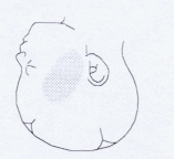
Figure 1. The optimal sensor placement on the fetal cheek
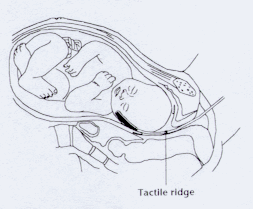
Figure 2. Intrauterine sensor placement.
Criteria considered after delivery :
-the ease of placing of the sensor and reception of a useable signal
-side effect either in the mother and in the fetus
- unpleasant or painful feelings of the mother
-the rate of Caesarian sections and spontaneous vaginal delivery placed into the context of suspicious and pathologic CTG-tracing
-mean FSpO2 by suspicious and pathologic CTG-tracing
-the newborns wellbeing was determined by the Apgar Score at 1st 5th and 10th min. and by postnatal arterial blood pH
-statistical analysis of gained data was anabled with the help of the following statistical tests:ANOVA or KRUSKAL-WALLIS
Results:
2. The method has proved to have no serious side effects neither on the mother nor on the fetus. No injury was caused to any mother due to the application and 30 minute placing of the sensor. If we do not consider occasional light impressions on the cheek at the fetus caused by the pressure of the sensor, these spontaneously disappearing in one hour after labor, we did not encounter any injury to the fetus which could be linked with the application of the sensor.
3. The feelings of the mother - the assessment of this indicated that there was one great fault from the perspective of the woman in labour and from the perspective of the obstetrician. Generally we can state that the application and presence of the sensor within the womans body is not painful but it provokes unpleasant feelings and also partially limits freedom of movement. During the uterine contractions, the patient does not percieve the presence of the sensor.
5. Average FSpO2 in percents (tab. 1, 2)
6. The status of a newborn (tab. 1, 2)
Table 1 schows the parametres of neonatal outcome in the year 1999.
|
|
||||||
|
|
|
|
|
|
||
|
|
|
|
|
|
|
|
|
|
|
|
|
|
|
|
|
|
|
|
|
|
|
|
|
|
|
|
|
|
|
|
Table 2 shows the parameters of neonatal outcome in the year 2000.
|
|
||||||
|
|
|
|
|
|
||
|
|
|
|
|
|
|
|
|
|
|
|
|
|
|
|
|
|
|
|
|
|
|
|
|
|
|
|
|
|
|
|
Altogether, IFPO does not involve any serious negative side effects. In the groups of suspicious CTG findings after evaluation of FSpO2 by means of IFPO there was no Ceasarean Section (CS) performed. However, a suspicious CTG curve is not a sufficient indication of CS, but still requires another examination and observation.
On the contrary, pathological CTG tracing is an absolute indication for labour termination. However, this group includes only 27 of labors terminated by CS after the verification of fetal status by FSpO2. FSpO2 significantly differed (p < 0.01 in the year 1999 and p = 0,01 in the year 2000) in the labors with pathological CTG tracing between fetuses delivered per sectionem and per vias naturales. Postportal states of newborns determined on the basis of their Apgar Scores in the 1st, 5th and 10th minutes, did not statistically differ. On the other hand, the states of newborns delivered vaginally and per SC, evaluated on the basis of umbilical arterial pH, differed significantly (p < 0.05 in the year 1999 only.) There was no statisticaly significant difference in umbilical cord pH in the cohort of patients examinated in the year 2000.
Discusion:
The results we gained are in agreement with published studies evaluating the influence of the evaluation of FSpO2 by means of IFPO on the frequency of acute performed CS. The study proved the method was easily feasible in common practical situations, without complications neither for the mother nor for the foetus. This method is more useful in the diagnosis of AIHP than pH examination from the head of the fetus and this is due to its easier applicability and noninvasivity.
If the IFPO is used in the diagnosis of AIHP, it is neccesary to understand its indications and rules during its usage. FSpO2 evaluation by means of IFPO is indicated in cases of suspicious or pathological CTG tracing testifying to imminent or starting fetal hypoxia. Severe and prolonged fetal bradycardia (with HR < 70/min and lasting longer than 5 minutes) indicated the necessity of immediate resuscitation of the fetus in utero ( acute tocolysis, O2) and immediate labor termination (CS, forceps). No delay of the birth by IFPO in these cases.
CTG monitoring remains the main screening method of AIHP. FSpO2 evaluation is an additional method of routine CTG examination. It is neither useful nor sufficient to monitore fetal state only by means of IFPO. If CTG tracing is physiological, there is no indication for IFPO moniotring. The exception is represented by an experimental study authorised by the Ethics Board, which evaluates FSpO2 during various situations and conditions [1]. If CTG tracing is suspicious or pathological the FSpO2 evaluation can make the situation clear or exclude these possibilities. If the FSpO2 >=30%, it can be assumed that in the present time the fetus is sufficiently supplied with oxygen. However a preexistent episode of acidosis as a result of hypoxia can not be excluded. In the case that FSpO2 >=30% a pH examination of the head of the fetus is not indicated. In the case of lasting suspicious or pathological CTG tracing, the following steps are recommended:
(1.) the repositioning of the woman giving birth to her left side and by this interrupting any possible compression of the vena cava inferior or the umbilical cord, (2.) correction of maternal hypotension and hydratation with coloids, especially in cases of epidural analgesia application, (3.) amnionfusion, (4.) a halt in the application of uterokinetics (oxytocin), (5.) acute tocolysis, (6.) applicatin of O2 to the mother by means of a mask.
In the case that FSpO2 >=30 %, the period of lowered saturation must last 10 minutes at least. It is recommended in this phase to follow the points mentioned above .
As soon as FSpO2 increases over 30 % or CTG tracing correct to a physiological curve either spontaneously or as a result of the points mentioned above, it can be supposed that in the phase considered fetal oxygenation is sufficient. Fetal state observation by means of CTG and IFPO could continue. In the case that FSpO2 remains under 30 % longer than 10 minutes, it is likely that the oxygen supply of the fetus is being harmed which can lead to hypoxic damage. If the clinic is not equipped with an appliance for the taking of blood samples from the fetal head to analyse blood gases, especially evaluating pH, the immediate termination of labor is recommended after previous resuscitation of the fetus in utero (acute tocolysis, O2 to mother). Where the evaluation of actual pH from the fetal head is possible, we can diagnostically use the modified Saling scheme. The level suspicious fetal hypoxia is determined at FSpO2 < 30 % for a period longer then 10 min. However, it is still not completely clear what the relation is between the percentual range of FSpO2 and the fetal hypoxia stage.
Animal studies offer some correlative schemes which are hardly applicable to human fetus. There have not been any sufficiently representative and interpretable human studies conducted in this area so far and that is mainly because of ethics problems. Along with the development of machine techniques concerning IFPO, it can be stated that there has been a definite lowering of median fetal oxygen saturation in both physiological and pathological labor. This was caused mainly by the lower calibration of old oxymetres for low FSpO2 in the course of labour [ 6, 11, 15, 21]. However, during physiological labour there is a gradual lowering of FSpO2. While in the cummulative phase of the 1st stage of labor the average FSpO2 of a non hypoxic foetus is 59 ± 10 %, in the 2nd stage of labour the levels of FSpO2 decrease slowly from 62 ± 9% (dilatation of the internal orifice of the cervix < 4cm), through 60 ± 11 % (5-7cm) orifice to 58 ± 10% before the disappearance of the internal orifice.
An often discussed question is the application of the sensor, the obtaining of a signal and its maintenance. The relatively mobile fetal head plays its role here, along with uterine contraction and the progression of the head down the birth canal. Repeated repositioning of the probe is necessary. In most cases it is not necessary to extract the probe and reinsert it. Usually, we do not have to be concerned about false result during any wrong application of the probe. In the case of the sensor being incorrectly placed and the signal not reaching a mininum of 50 %, the FSpO2 cannot be evaluated ( the display does not show any level). The turbid fetal fluid with meconium does not influence the reliability of obtain values. However the obtained of a quality signal can be more difficult [20].
Despite the fact that the development of probes continues the contemporary method of plaining the probe freely between the cheek and uterine wall seems to be the most useful. Probes with the original design, whose positioning was based on the principle of scalps CTG electrodes, have not proved successful. Among the reasons for this are: traumatisation with possible excoriation of presenting part of the fetus as well as non-representative result obtained from often ischemished obstetrical injuries. Contemporary probes have not shown any negative effects [3, 9, 10, 11, 14], which was confirmed in this study. There are interesting findings with very good correlations between FSpO2 and pH in the umbilical artery. The results of both methods show very high predicted values for the postpartal states of fetuses and this is in the case of both physiological and pathologic labor. The easier method of application of the sensor is during epidural analgesia because of the lowered perceptivity of tactile stimules from the birth canal. The positive influence of epidural analgesia on the values of FSpO2 has been proved in the course of physiological labour.
Eventhough there have been many coinducted studies concerning the influence of IFPO on the frequency of acute obstetrical operative interventions, the majority of them partially invalidated by small number faults or methodological problems. Although several multicentric studies show some trends, a definitive evaluation of IFPO in this area of obstetrics has not been released yet. The Czech Republic is still waiting for the conducting of a prospective controlled study.
Despite the lack of definite and clear conclusions, it can be stated that the intrapartal evaluation of FSpO2 by means of IFPO for fetus with suspicious or pathological CTG tracings leads to more precise diagnosis of acute fetal hypoxia and the lowering of acute CS rate performed due to this indication.
References: